Friday, April 16, 2010

Opportunist

  • cause disease in people who are immunocompromised
Endogenous opportunist
Candida albicans
Morphology: yeast(blastospore), pseudohyphae(like links), true hyphae (have septa, paralel walls)


Candida albicans in rabbit kidney
very light stain

Candida albican in liver


Candida albican
this is a culture (most likely not tested)
Candida albican Chlamydospores
in starvation medium "corn meal agar test"
Candida albicans in sputum
over decolorized slide
Rhizopus in tissue
wide nonseptate hyphae
irregualr branching (90 deg)
Rhizopus in kidney
wide nonseptate hyphae
irregular branching
Pneumocystis carinii
cause pneumonia in AIDS patients
note the brown red blood cells
Pneumocystis carinii
Cryptocossus neoformans in tissue
has fuzzy coloring capsule
barrow base buddings
Extracellular
Mating type a & alpha
haploid dominant
Serotype: D (found in Europe) associate with AIDS& children in NYC

Friday, April 9, 2010

Systemic fungi

  • normal habitat: soil
  • can cause pulmonary infection when inhaled
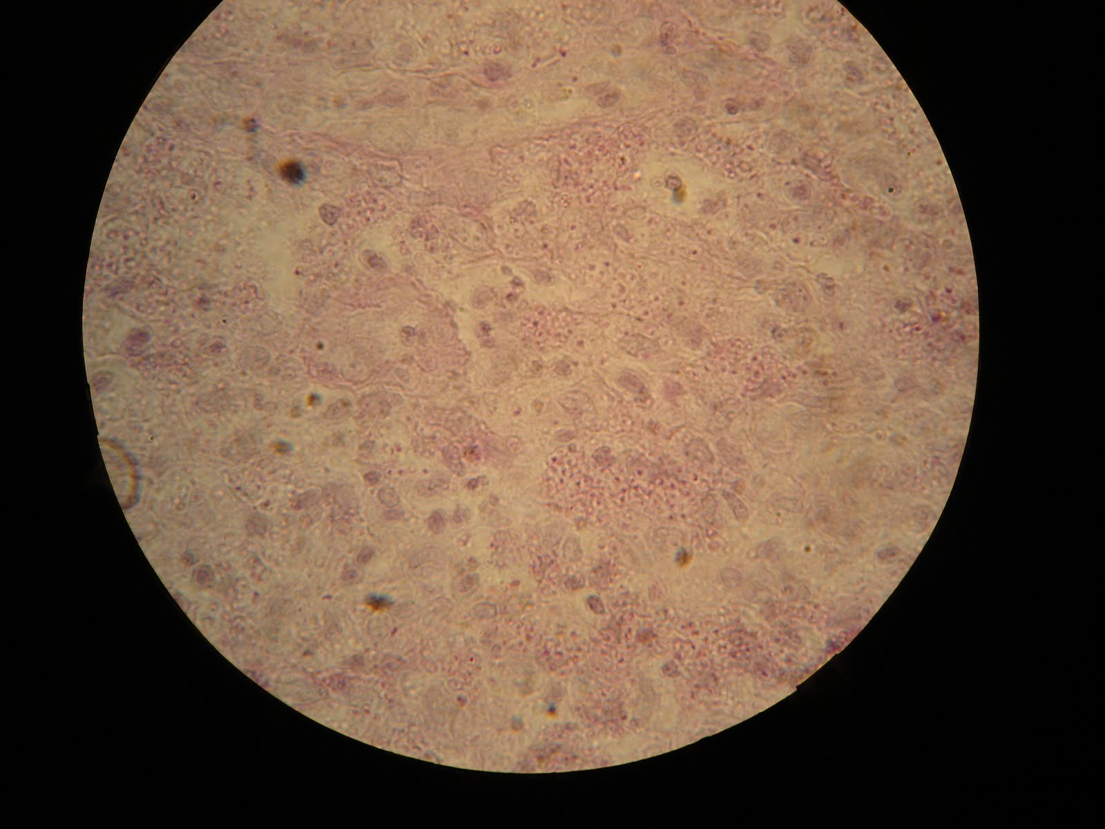
Blastomyces dermatitidis
Blastomyces dermatitidis tissue section

Blastomyces dermatidis mycelial phase
Conidia wider than pointer 4-5 micrometer

Blastomyces dermatitidis yeast phase (lactophenol blue wet mount)
Yeast wider than pointer 8-20 micrometer
Blastomyces dermatitidis tissue section (Gomori silver stain)
yeast wider than pointer (8-20 micrometer)

Histoplasma capsulatum
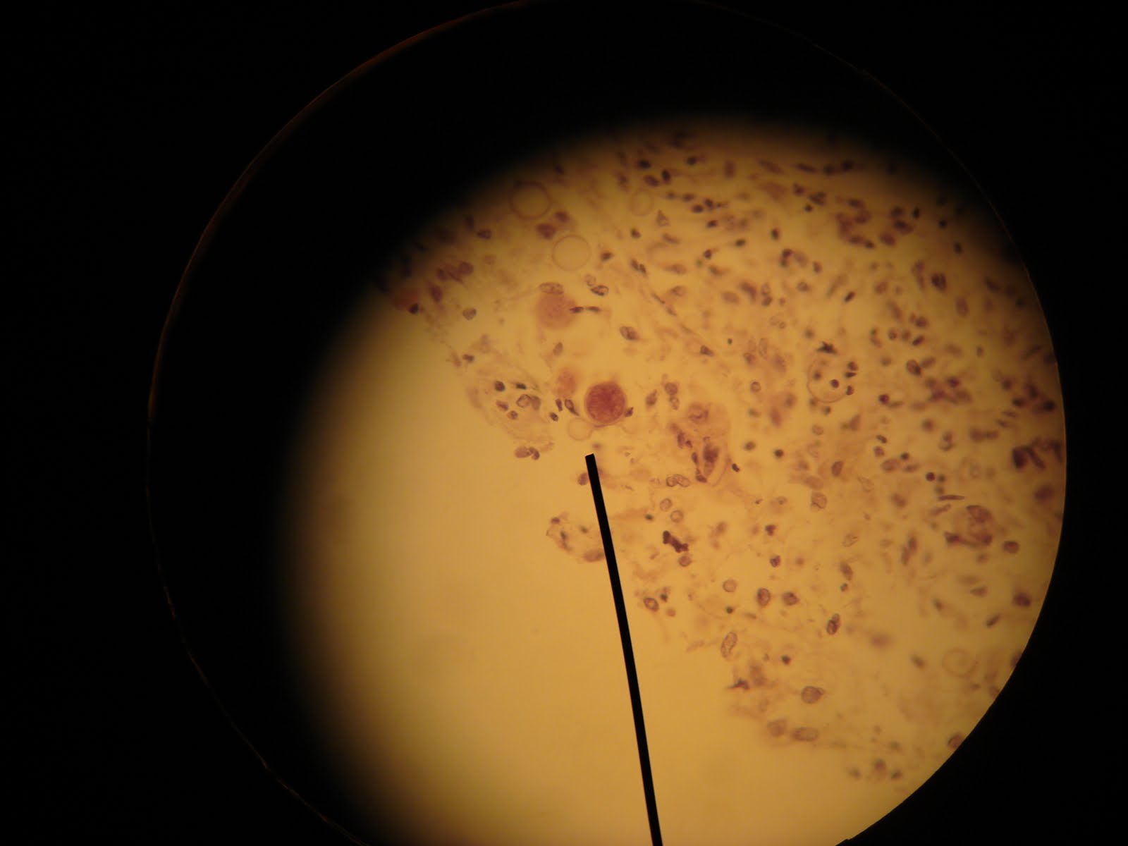
Histoplasma capsulation in brain tissue (Gomoro stain)
Magnification 100X
Histoplasma capsulatum in tissue
100X


Histoplasma capsulatum mycelial phase (slide culture wet mount)
8-20micrometer, 400X
note the spiky conidia

Paracoccidioides brasiliensis 
No sexual phase
37 deg C: multiple buds yeast with narrow bases (multinucleate, need sulfur containing aminoa acid)
Disease: Lesions [due to conidia inhalation]
Subclinical (no symptom just flu, nodules & plaques)
Chronic unifocal (adult smoker, whose 1 lung infected)
Chronic multifocal (resemble TB, cough, chest pain, weight loss)
Subjuvenile
AIDS

Distribution:
South, Central America
humid Summer & dry Winter
Rural>>Urban
Clinical disease M>>F
-Estrogen inhibit tranformation of Y to H
-Estrogen bing protein found in cytoplasm

Diagnose:
Skin test
Serological test

 Paracoccidioides brasiliensis yeast phase (lactophenol blue wet mount)
10-60 micrometer
multiple buds with narrow bases
Paracoccidiodes brasiliensis tissue section (Gomori stain)
multiple buds with narrow bases
Agents that cause Mycetoma
granule from Mycetoma (Madura foot)
causative agent: fungus

Coccidiodes immitis
"immitis" = not mild, severe biohazard
18S rRNA
25 deg C: septate hyphae & alternating arthroconidia with disjunctions that lyse [saprophytic that ruptures with wind]
37 deg C: slimy polysaccharide, yeast with large endospores [in host]

Habitat: alkali soil, arid climate (southern Arizona, central California, Southern New Mexico, & west Texas)

Disease: MOST DANGEROUS SYSTEMIC MYCOSES
Acute Coccidiodomycetoma [inhaling the arthroconidia, effect ppl with low immune system]
-endospores germinate in the lungs (rare, but lethal)
-erythema nodusum (red infected lumps)
-Adult M> Adult F (hormomal control)
Chronic cutaneous Coccidiodomycosis
-High risk for: HIV patients, organ transplant, pregnant woman (hormone)

Diagnose:
Skin test
Serological test
Microscopy


Coccidiodes immitis sperule
note the large endospores

Coccidiodes immitis sperule in human lung tissue (Gomori silver stain)
humungus endospores


Coccidioides immitis sperule (KOH mount)
spot the endospore

Artifact: this is fat globule that can be mistaken for sperule or yeast.

Geotrichum mycelia (lactophenol blue wet mount)

Coccidiodes immitis mycelial phase (Lactophenol blue wet mount)
Sporothrix schenckii
  • dimorphic
  • 22 deg C: fine septate hyphae with cluster of conidia
  • 37deg C: elongated yeast cells with cigar-shaped buds (the form when they infect tissues)
Habitat: soil, animal excreta, living or decaying vegetative
Disease: Sporotrichosis [caused by animal bites, rose thorns, insect sting...]
-papule enlarges overdays or weeks
-can persist for years
-does not cure without treatment
-erythematous(redness from inflamation), ulcerated, pus

Sporothrix schenckii yeast phase from a laboratory mouse
smaller than pointer 1-3 micrometer
look for cigar shaped yeast
Sporothrix schenckii yeast phase lactophenol blue wet mount
look for cigar shaped yeast
Sporothrix schenckii mycelial phase (stain of slide culture)
very fine, delicate
smaller than pointer

...

Scopulariopsis spp.geicohands-like conidiophore
large, rough, annelloconidia (produced by annelides)
Aspergillus spp.
Conidiophores with phialoconidia
brush like
similar with Penicillium but has more volume & shorter conidia
Superficial Mycoses
  1. Disease:
  • Pityriasis versicolor (patches altered pigment, versicolor & scaling)
  • Seborrhoeic dermatitis (includes Follicular pityriasis & Dandruff)


causative agent: Malassezia furfur
hyphae & yeast (spaghetti & meat balls)
Basidiomycete
Lipophilic yeast (loves fat/lipid)
can be systemic => nutritional, supplementation, associate with catheters  

  1. Tinea nigra
      • causative agent: Exophiala wernekii (dimorphic, Ascomycete, yeast in culture; mold in lession)
      • form brown to black spots
      • tropical disease
      • palm & soles
      • young F>M 
  2. Piedra: infect hair, formation of waxy small, firm masses on hair
    • Black
      • black nodules on scalp
      • causative agent: Piedraia hortae ()
      • tropical, M>F (swimmers)
    • White
      • white nodules on beard, mustache, genital region
      • causative agent: Trichosporon beigelii (mold, septate, hyphae)
      • temperate & tropical, south US, M>F
Dermatophytes (skin infection)
  • Trichophyton
  • Microsporum
  • Edidermophyton
All molds, septate hyphae
30-70% adults
incident increase with age

Monday, April 5, 2010

Techniques, Sapprophyte

Sexual stages of 3 classes:

Basidiomycetes
brown basidiospore


Zygomycetes
dark colored zygospore formed from fusion of 2 aseptate hyphae


Ascomycetes
the 2 celled ascospores are produced in a sac like ascus

Dermatophytes and other fungi that causes superficial infections:
limited to keratinized tissues. ie. skin, hair, nails
dermatophyte infection determined by observing skin scrapping in KOH wet mount
has macroconidia & microconidia


Microsporum canis
spindle-shaped, thick walled macroconidia.
has up to 15 septa.

Microsporum gypseum
thin wall
usually 4-6 septa

Epidermophyton floccosum
club-shaped macroconidia
2-3 septa
no microconidia


Tricophyton mentagrophytes
cigar-shaped
thin walled macroconidia
abundant
coiled hyphae
hair perforation positive


Tricophyton rubrum
cigar-shaped macroconidia
thin walled microconidia
Dermatophyte
anthropophillic (attracted to man*), zoophillic, geophillic [as pop. decrease, lose ability to mate]

Saprophytes: Organisms that live on dead organic matter


Aspergillus
chained conidia on phialides arise from swollen vesicle
Exogenous opportunistic fungus.


Scopulariopsis spp.
geicohands-like conidiophore
large, rough, annelloconidia (produced by annelides)


Penicillium
chains of conidia from branched conidiaphore
brush like, longer than Aspergillus

Penicillium marnefei
severe biohazard
intracellular (Histo too!!)
dimorphic
haploid, no sexual stage
most infected by HIV positive patients

Disease:(by inhalation)
once infected, cannot be cured
effect immune surpress individuals (ie. HIV)

Disribution:
Southeast Asia, nothern Thailand
Diagnose:
Culture, PCR, Serological test


Rhizopus
non-septate, wide hyphae
sporangia filled with sporangiospores
has rhizoids at base of sporangiophore
Rndogenous opportunistic fungi

Mucor
septate hyphae
sporangiospores in sporangia.

Alternaria
dematiaceous (dark colored)
club-shaped conidia